Brain cancer prediction has reached new heights with the advent of advanced artificial intelligence, providing hope for more accurate assessments of pediatric glioma patients. A recent study by researchers at Mass General Brigham has showcased a cutting-edge AI tool that excels in predicting the relapse risk of brain cancer in children by analyzing multiple MRI scans over time. This innovative approach, which utilizes temporal learning in AI, surpasses traditional methods, offering improved insights into the likelihood of recurrence for young patients. By integrating AI cancer detection with comprehensive MRI scan analysis, healthcare professionals can now better understand and anticipate the complexities of childhood brain tumors. Enhanced predictions not only alleviate the emotional burden on families but also pave the way for personalized treatment plans that could significantly improve outcomes for children facing this daunting diagnosis.
The landscape of predicting brain tumors, particularly in young patients, is undergoing a transformative shift, driven by breakthroughs in artificial intelligence and imaging techniques. In the realm of pediatric oncology, the ability to assess the risk of pediatric glioma recurrence is vital for timely interventions. Innovative methodologies are being embraced, enhancing the way clinicians utilize MRI analysis to monitor changes in tumor behavior over time. As we delve into these advancements in brain cancer assessment, the focus on AI-driven solutions underscores the potential for improved patient care and the vital role of modern technology in identifying relapse risks effectively.
Understanding Pediatric Glioma and Its Challenges
Pediatric gliomas are a diverse group of brain tumors that primarily affect children and are categorized based on the cell type and location within the brain. These tumors can vary significantly in behavior and treatment responses, making their management particularly challenging. While many pediatric gliomas are treatable with surgery alone, the likelihood of recurrence remains a top concern for healthcare providers and families alike. The need for effective monitoring and early detection systems is crucial to ensure children receive timely and appropriate interventions.
Understanding the unique characteristics of pediatric gliomas can aid in developing better predictive models for relapse. Factors such as tumor grade, location, and age at diagnosis can influence how aggressive the treatment needs to be and how closely patients should be monitored. Advances in imaging technology and AI-based systems are paving the way for innovative strategies to predict recurrence, allowing for a more tailored approach to care.
AI Cancer Detection: Revolutionizing Brain Tumor Monitoring
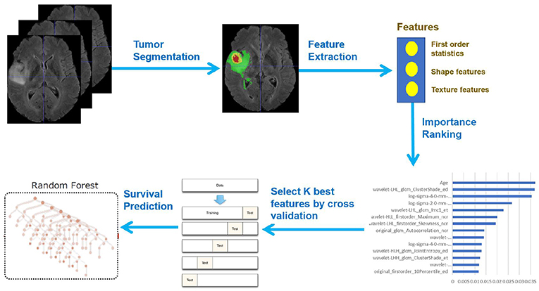
The integration of artificial intelligence (AI) in cancer detection represents a significant milestone in medical research and patient care. In the realm of pediatric gliomas, AI tools enhance the accuracy of monitoring methods by analyzing vast datasets from brain scans. Traditional imaging techniques often fall short in predicting patient outcomes, but AI-driven models offer a sophisticated analysis that can uncover patterns in imaging that human eyes might miss.
By harnessing algorithms that can process thousands of MRI scans, researchers can develop models that not only identify existing tumors but also predict the likelihood of recurrence. The study illustrated in the recent research from Mass General Brigham demonstrates how AI cancer detection can significantly outperform traditional methods, offering hope for more effective and less invasive monitoring strategies for children diagnosed with brain tumors.
Frequently Asked Questions
How does AI cancer detection improve brain cancer prediction in pediatric patients?
AI cancer detection enhances brain cancer prediction in pediatric patients by analyzing multiple MRI scans over time, allowing for more accurate assessments of relapse risk. Traditional methods often rely on single scans, which limits their predictive accuracy. The advanced algorithms can identify subtle changes over consecutive scans, significantly improving the prediction of outcomes for conditions like pediatric glioma.
What is temporal learning in AI and how does it relate to brain cancer prediction?
Temporal learning in AI refers to a technique that analyzes the sequence of MRI scans taken over time to identify patterns and changes that may indicate cancer recurrence. In the context of brain cancer prediction, especially for pediatric glioma, this method enables AI models to improve accuracy by learning from temporal data rather than relying solely on isolated scans.
What are the main benefits of using MRI scan analysis for brain cancer prediction?
The main benefits of using MRI scan analysis for brain cancer prediction include enhanced detection of tumor changes, improved accuracy in assessing relapse risk, and the ability to tailor patient monitoring and treatment strategies based on individualized risk profiles, particularly for pediatric glioma cases.
How accurate is AI in predicting relapse risk for pediatric glioma compared to traditional methods?
AI-driven models have shown to predict the relapse risk for pediatric glioma with an accuracy of 75-89%, significantly outpacing the approximately 50% accuracy of traditional methods that rely on single MRI scans. This heightened precision can lead to better treatment decisions and outcomes for young patients.
What future developments can we expect in brain cancer prediction through AI technology?
Future developments in brain cancer prediction through AI technology may include the expansion of clinical trials to validate AI-informed risk predictions, the integration of temporal learning techniques across different medical imaging scenarios, and potential enhancements in treatment protocols based on individualized relapse risk assessments for patients with pediatric glioma.
Can AI tools help reduce the burden of frequent MRI scans for pediatric glioma patients?
Yes, AI tools can potentially reduce the burden of frequent MRI scans for pediatric glioma patients by accurately identifying low-risk individuals. By applying advanced prediction models, clinicians may be able to decrease the monitoring frequency for these patients, thereby minimizing stress and discomfort associated with repeated imaging.
What role does the National Institutes of Health play in brain cancer prediction research?
The National Institutes of Health (NIH) plays a critical role in brain cancer prediction research by funding studies that explore innovative AI technologies and their application in medical settings. Their support helps facilitate research collaborations and advancements in predictive modeling for pediatric conditions like glioma, ultimately aiming to improve patient care.
| Key Points | Details |
|---|---|
| AI Tool for Prediction | An AI tool outperforms traditional methods in predicting relapse risk in pediatric brain cancer. |
| Study Overview | Conducted by researchers at Mass General Brigham and published in a reputable journal, it analyzed nearly 4,000 MR scans. |
| Temporal Learning Technique | This approach synthesizes multiple brain scans over time to improve prediction accuracy. |
| Prediction Accuracy | The AI model achieved a 75-89% accuracy rate in predicting glioma recurrence, surpassing traditional methods. |
| Future Implications | The researchers aim to conduct clinical trials to validate this AI tool’s effectiveness and enhance pediatric treatment. |
Summary
Brain cancer prediction has made significant advances with the development of AI tools that analyze brain scans to better predict relapse risks in pediatric patients. The recent study demonstrates that these AI systems can provide more accurate assessments than traditional imaging techniques, enabling earlier identification of high-risk patients. By leveraging temporal learning techniques, researchers expect to improve personalized treatment strategies, ultimately leading to better outcomes for children facing brain cancer.